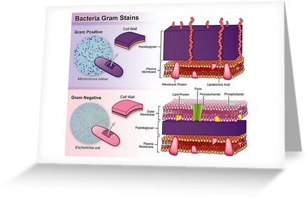

Gram negative cells have an outer membrane that resembles the phospholipid bilayer of the cell membrane. Gram positive bacteria also have teichoic acids, whereas Gram negatives do not. Gram positive cells have thick layers of a peptidoglycan (a carbohydrate) in their cell walls Gram negative bacteria have very little. Gram stain results reflect differences in cell wall composition.
#Gram positive vs gram negative color professional
Knowing the Gram reaction of a clinical isolate can help the health care professional make a diagnosis and choose the appropriate antibiotic for treatment. Since then, the Gram stain procedure has been widely used by microbiologists everywhere to obtain important information about the bacterial species they are working with. This very commonly used staining procedure was first developed by the Danish bacteriologist Hans Christian Gram in 1882 (published in 1884) while working with tissue samples from the lungs of patients who had died from pneumonia.
#Gram positive vs gram negative color how to
You will learn how to prepare bacterial cells for staining, and learn about the gram staining technique. Some examples of differential stains are the Gram stain, acid-fast stain, and endospore stain. Differential stains use more than one stain, and cells will have a different appearance based on their chemical or structural properties. Scientists will often choose to perform a differential stain, as this allows them to gather additional information about the bacteria they are working with. Isolated and imaged by Muntasir Alam, University of Dhaka, Department of Microbiology in 2007. Image 1: Microscopic view of Bacillus (rod) shaped bacteria simple stained with crystal violet. Basic stains, having a positive charge, bind strongly to negatively charged cell components such as bacterial nucleic acids and cell walls. The single dye used here in our lab is methylene blue, a basic stain. In a simple stain, a bacterial smear is stained with a solution of a single dye that stains all cells the same color without differentiation of cell types or structures. The purpose of staining is to increase the contrast between the organisms and the background so that they are more readily seen in the light microscope. Living bacteria are almost colorless, and do not present sufficient contrast with the water in which they are suspended to be clearly visible. Simple stains can be used to determine a bacterial species’ morphology and arrangement, but they do not give any additional information. Some stains commonly used for simple staining include crystal violet, safranin, and methylene blue. One type of staining procedure that can be used is the simple stain, in which only one stain is used, and all types of bacteria appear as the color of that stain when viewed under the microscope. Members of the phyla Proteobacteria (exception: some members of the order Rickettsiales), Cyanobacteria and Spirochaetota.\) Members of the phyla Bacillota and Actinomycetota (exception: genus Mycobacterium). Remove the excess of fluid with a paper towel and allow to air dry until the specimen is completely dry.Flood gently with acetone-ethanol solution.Add Lugol's solution (which contains iodine), wait for 1 minute.Add chrystal violet, wait for 1 minute.


 0 kommentar(er)
